


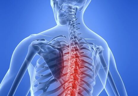
Osteochondrosis - refers to diseases based on a degenerative dystrophic process affecting the intervertebral discs as well as other structural elements of the spine: vertebral bodies, intervertebral joints, ligaments, tendons.
Thoracic vertebral osteochondrosis is a rare pathological form. This is due to the peculiarities of the anatomy of the upper part of the bone. The thoracic spine is made up of 12 vertebrae that are connected to the ribs, the front of which is connected to the sternum. Rugged Frame - Chest protects vital organs (heart, lungs) from injury.
This bone structure not only limits the mobility of this part of the spine, but also protects it from the negative effects of physical activity, as well as the premature destruction of the intervertebral discs.
The intervertebral disc is the cartilage layer between the vertebrae and consists of the central part - the gelatinous nucleus pulposus and the annulus fibrosus - the capsule.
The intervertebral discs provide stability of the spine against vertical loads, act as shock absorbers when walking, running, jumping, and together with the other joints of the vertebrae, provide mobility and flexibility of the spine.
Development of thoracic osteochondrosis
In osteochondrosis, the blood supply deteriorates and the transport of water, glucose, and amino acids to the nucleus pulposus, which is necessary for the synthesis of water-bound carbohydrates, is disrupted. The core dries out and the gel-like structure becomes fibrous, thus losing its elasticity and ability to dampen shocks. The load falls on the injured annulus and vertebrae. Microcracks appear on the annulus fibrosus, the fibers are stretched, and the nucleus pulposus can no longer be fixed, and the nucleus pulposus begins to protrude into the spinal canal - a herniated disc. When the annulus fibrosus ruptures, an intervertebral hernia forms.
cause of disease
Osteochondrosis of the thoracic spine develops as a result of the body's natural aging in people over 40-45 years of age. This is manifested as a slowing of the regeneration process of cartilage and bone tissue, a reduction in the production of collagen, thus maintaining the elasticity and strength of the spinal ligaments.
At a young age, the rapid progression of osteochondrosis in the thoracic region occurs against a background of pathology that negatively affects the condition of the cartilage and bone tissue of the spine.
- Systemic connective tissue diseases: rheumatoid arthritis, scleroderma.
- Endocrine diseases: diabetes, hypothyroidism.
- Congenital and acquired postural abnormalities: kyphosis, scoliosis.
- Long-term exposure to static and dynamic loads.
- Genetic predisposition to cartilage weakness.
- Thoracic spine trauma.
A sedentary lifestyle, an unhealthy diet, obesity, and a lack of vitamins and trace elements in the body can lead to premature disc damage.
degree of pathology
The greater the deformation of the intervertebral discs and vertebrae, the more obvious the clinical manifestations.
Stages of disc destruction in thoracic osteochondrosis:
I'm on stage. Because the nucleus pulposus cannot retain the water it needs to restore its tissue, the disc begins to gradually collapse. The fibrous annulus is full of cracks. Patients experience periodic chest discomfort following physical exertion.
Stage two. The disc continues to be destroyed, the annulus fibrosus fibers are delaminated, and the nucleus pulposus migrates into the deep fissure formed on the surface of the disc. The height of the intervertebral disc decreases and the mobility of the vertebrae increases. The back muscles in the damaged segmental area tense reflexively in an attempt to limit mobility in the thoracic area. Moderate pain.
The third phase. If the integrity of the annulus fibrosus is compromised, the nucleus pulposus can enter the spinal canal, forming an intervertebral herniation. Compression of spinal cord structures: nerve fibers, blood vessels. The vertebral bodies were also deformed, and bone tissue growth in the form of osteophytes was observed. The pain becomes constant and the range of motion of the thoracic spine decreases.
Phase IV. In the final stages of thoracic osteochondrosis, signs of a degenerative process are observed on the ligaments, muscles, and other tissues surrounding the affected spinal segment. The cartilage of the disc is replaced by scar tissue. Osteoarthritis occurs in other vertebral joints. The clinical presentation is variable and depends on the extent of the disc injury and the location of the hernia.
If spinal cord compression occurs, radiculopathy, myelopathy, and other irreversible consequences can result in disability.
If the problematic disc is covered with fibrous tissue, the adjacent vertebrae fuse, which can move the disease into stable remission, but with a loss of function of part of the spine, becoming immobile in the area of the affected part.
Phase IV. This is the final stage of the disease. The cartilage of the intervertebral disc is replaced by connective tissue, and adjacent segments of the spine are involved in the pathological process. The joints grow together and become immobile (stiff). The patient is seriously ill: not only severe pain in the neck, but also severe pain between the arms, chest, and shoulder blades, signs of cerebrovascular accident, and sensitivity disorders. This is a life-threatening condition that can lead to a stroke.
The success of treatment is 90% dependent on the experience and qualifications of the physician.
Doctor free consultation and diagnosis
- Chiropractor
- Vertebrate scientist
- Osteopath
- neurologist
Perform a thorough diagnosis of the entire spine and each segment in consultation with a doctor. Doctors determine which segments and nerve roots are involved and cause painful symptoms. Based on the results of the consultation, detailed treatment recommendations are made and additional diagnoses are made if necessary.
Signs and symptoms of thoracic osteochondrosis
Symptoms of thoracic osteochondrosis are often mistaken for clinical manifestations of other disorders. This is because when the spinal roots are compressed, the function of the organs they innervate is disturbed. The gastrointestinal tract, liver, pancreas, heart are not working properly.
The pain in the chest is not obvious, but can occur in the arms, ribs, collarbone, shoulder blades, and abdomen. Due to the nature of osteochondrosis pain, they resemble episodes of angina, acute pancreatitis, or cholecystitis.
Often, pain between the shoulder blades is accompanied by a feeling of lack of air, which many believe is a heart attack.
With significant and prolonged compression of the spinal cord root, severe neuropathology develops into motor and sensory impairments. Specifically, the localization of the disease depends on which thoracic vertebra is near the nerve root.
Areas of pain and sensitivity changes in the form of numbness extend from the neck, shoulder blades, ribs, sternum to the abdomen.
Principles of Disease Diagnosis
The diagnosis of osteochondrosis involves the following steps:
- Collect medical records.
- Clinical examination to assess neurological status.
- function test.
- Instrumental Methods: X-ray, Magnetic Resonance and Computed Tomography.
An important stage of examination is differential diagnosis. Symptoms of thoracic vertebral osteochondrosis are often "camouflaged" as heart, stomach, and lung disease, so additional research methods are needed to make a proper diagnosis.
treat
Most patients with signs of thoracic osteochondrosis require conservative management. Surgical treatment is performed only in particularly severe cases, where the spinal canal is significantly narrowed by the hernia and the spinal cord is severely compressed.
In modern clinics for the treatment of osteochondrosis, the use of the non-surgical author's method not only eliminates pain in the acute phase, but also stabilizes the condition of the spine and prevents complications. For each patient, a treatment strategy was chosen based on the severity of the pathology.
Osteochondrosis of the thoracic spine: symptoms and treatment of the modern clinical spine
Goals of Osteochondrosis Pharmacotherapy:
- Block pain syndrome.
- Reduce inflammation.
- Normalizes metabolic processes.
- Improve blood supply.
- Relieve muscle spasms.
Medications used: Anesthetics, anti-inflammatory drugs, steroids, muscle relaxants, B vitamins.
Modern medical centers have improved traditional manual therapy methods, adding electrophoresis and photodynamic laser therapy to improve the therapeutic effect.
Treatment includes:
- Soft manual technique that works on a physiological level, allowing you to successfully eliminate pinched nerve roots in the spine.
- Multicomponent electrophoresis is a medical procedure in which drugs are delivered directly into the lesion.
- Laser Treatment. Under the action of laser radiation, drugs applied to the skin of the affected segment of the spine can penetrate 10-15 cm deep and have analgesic and anti-inflammatory effects at the cellular level.
A paravertebral block is a method of introducing anesthetic into the area of a damaged nerve root to help quickly relieve pain, reduce swelling, inflammation, and improve blood supply.
Shockwave therapy, where specific frequencies of acoustic vibrations create an effect similar to a power massage. The therapeutic effect of this procedure lies in analgesic effect and enhanced tissue regeneration.
Physical therapy exercises that strengthen the back muscles help create a naturally strong corset that holds the spine in the correct anatomical position.
Years of experience in the treatment of osteochondrosis of the thoracic spine in specialized clinics have shown that with a correct and comprehensive treatment, the symptoms that complicate the life of the patient disappear, thus preventing the further development of the pathological process.